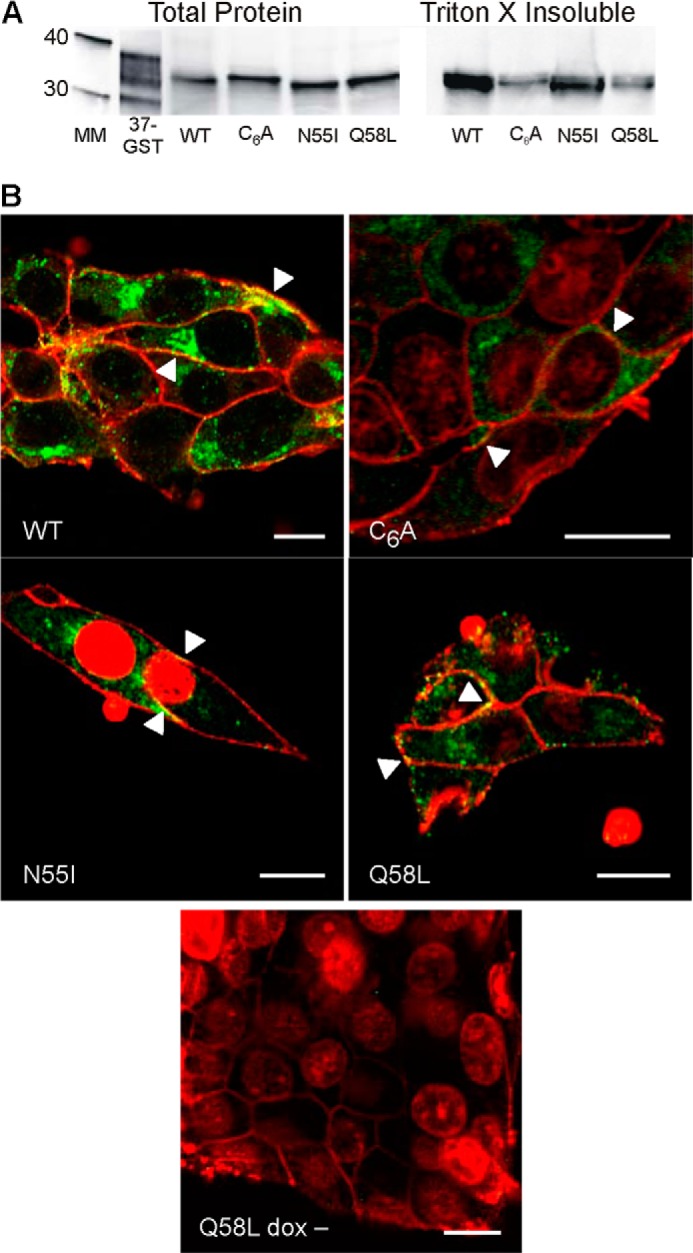
FIGURE 1.

Expression and localization of Cx37 are comparable in Cx37-WT, -C6A, -N55I, and -Q58L iRin cells. A, Cx37 was detected in both the whole cell and Triton X-insoluble protein fractions of iRin cells expressing Cx37-WT, -C6A, -N55I, and -Q58L; 50 μg of total protein was loaded for each cell type. The 37-GST (GST-rCx37CT229–333, where CT indicates C terminus) lane shows positive control for antibody detection; note that the Cx37-GST fusion protein typically migrates as multiple bands (this lane was contrast-enhanced for better band visibility) (7, 9). Positions of the 30- and 40-kDa molecular mass markers are shown in the mass marker (MM) lane. B, immunocytochemistry revealed localization of WT and mutant Cx37 (green) at appositional and non-appositional membranes (arrowheads). Red staining corresponds to biotinylated proteins on the extracellular surface of the plasma membrane and to ToPro3-labeled nuclei in the C6A, N55I and Q58L images (some ToPro3-labeled nuclei are evident in unattached cells above the plane of focus). Yellow staining corresponds to Cx37 localized to the plasma membrane. Scale bars: 10 μm.
