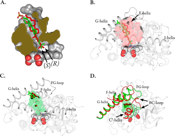
FIGURE 4.
A, slab view of the CYP142A2 (PDB code 4TRI) active site complexed with cholesteryl sulfate (green sticks). The CYP125A1 (red, PDB code 2X5W) and CYP142A2 (white, PDB code 2YOO) 4-cholestene-3-one ligands. B, the CYP125A1 active site in complex with 4-cholestene-3-one (red sticks) overlaid with the CYP142A2 cholesteryl sulfate ligand (green). The inner lining of the binding pocket has been colored semi-transparent red. C, the CYP142A2 active site complexed with cholesteryl sulfate (green sticks) overlaid with the CYP125A1 4-cholestene-3-one ligand in red. The inner lining of the binding pocket has been colored semi-transparent green. D, CYP142A2 with the FG and BC helices colored green. The corresponding regions of CYP125A1 have been superimposed in red.

