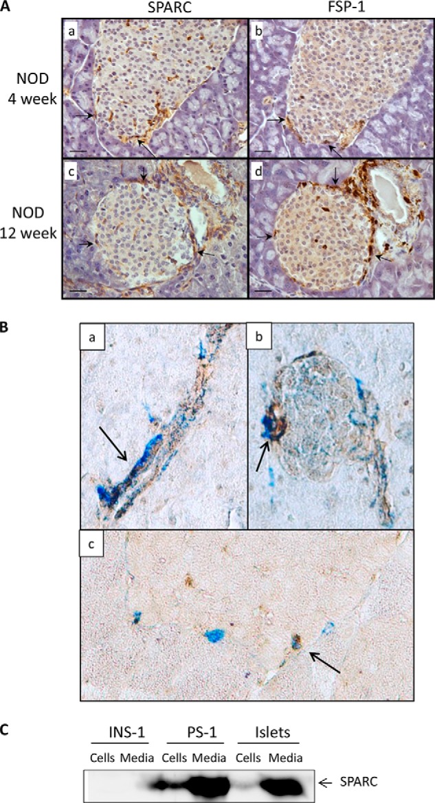
FIGURE 2.

SPARC is expressed by stromal cells within islets. A, adjacent sections (5 μm) of 4- and 12-week NOD mouse pancreas were stained with antibodies to SPARC (left panels) and FSP-1 (right panels) followed by biotin-conjugated secondary antibody. Positive areas were visualized using the ABC/DAB method producing a brown color. Representative images are shown of multiple sections analyzed from at least three different mice. Arrowheads indicate examples of SPARC and FSP-1-positive cells. Scale bar, 20 μm. B, pancreatic sections from ICR mice were co-stained with antibodies to SPARC (brown DAB substrate) and Fsp-1 (Vector Blue substrate). Representative images of staining in ducts and islets are shown, taken with 20× objective and with additional 2× digital zoom in panels a and c. Arrows indicate areas of co-staining. In C, concentrated medium and cell lysates from equal numbers of INS-1 and PS-1 cells, or from intact islets (groups of 50 islets) were analyzed by Western blot and probed with antibodies to SPARC.
