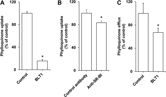
FIGURE 3.

Effect of SR-BI inhibitors on vitamin K1 apical transport by differentiated Caco-2 TC-7 monolayers. A, effect of BLT1 on vitamin K1 uptake. The apical sides of the cell monolayers were preincubated for 60 min with either BLT1 at 10 μm or DMSO (control), before receiving FBS-free medium containing phylloquinone-enriched mixed micelles at a 2.5 μm concentration and supplemented with either BLT1 at 10 μm or DMSO. The basolateral sides received FBS-free medium. Incubation time was 60 min. Data are means ± S.E. (error bars) of three assays. *, significant difference with the control. B, effect of SR-BI-blocking antibody on vitamin K1 uptake. The apical side of the cell monolayers was pre-incubated for 5 min with either 3.75 μg/ml of anti-human SR-BI antibody or 3.75 μg/ml of anti-human CD13 antibody (control), before receiving FBS-free medium containing phylloquinone-enriched mixed micelles at a 2.5 μm concentration. The basolateral side received FBS-free medium. Incubation time was 60 min. Data are means ± S.E. of 3 assays. *, significant difference from the control. C, effect of BLT1 on vitamin K1 apical efflux. Cell monolayers were first enriched in phylloquinone during 4 h. The apical side of the monolayers was then carefully rinsed and received either FBS-free medium containing vitamin K1-free mixed micelles or the same mixture plus BLT1. The basolateral side received FBS-free medium. Efflux time was 60 min. Data are means ± S.E. of three assays. *, significant difference from the control (assay performed without BLT1).
