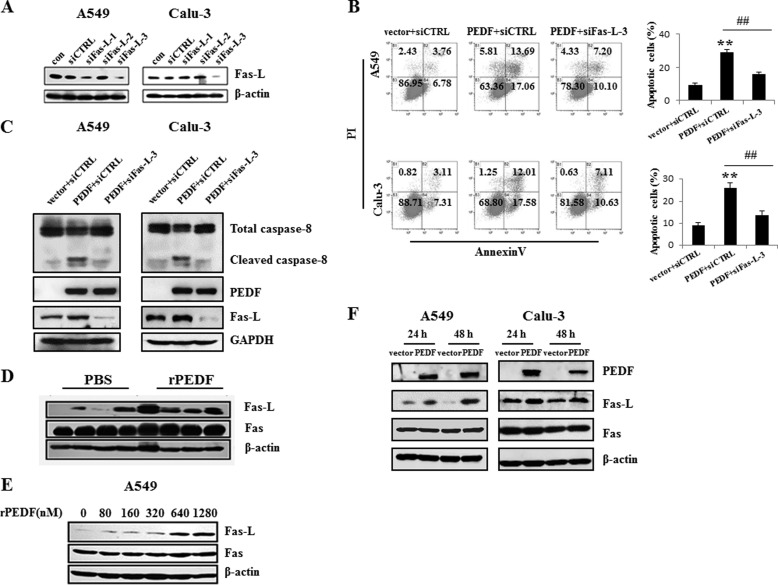
FIGURE 5.
PEDF-induced apoptosis is Fas-L-dependent. A–C, after 48 h, the interference effects of the Fas-L siRNAs (siFas-L-1/2/3) were analyzed by Western blot analysis. Fas-L siRNA-3 (siFas-L-3) or nonspecific siRNA was transfected into A549 and Calu-3 cells for 12 h before PEDF gene or vector transduction. After 48 h of serum depletion, the cells were harvested for the quantification of the apoptotic cells by flow cytometry (B) and Western blot analysis (C). Representative diagrams are shown. The apoptosis results are presented as mean ± S.D. of three independent experiments. **, p < 0.01 versus vector + siCTRL-treated cells; ##, p < 0.01 versus PEDF + siCTRL-treated cells. con, control; siCTRL, control siRNA; PI, propidium iodide. D–F, the protein levels of Fas-L, but not that of Fas, were up-regulated in the tumor tissues (first through fourth lanes, from PBS-treated mice; fifth through eighth lanes, from rPEDF-treated mice) (D) or A549 cells (E) by rPEDF in a dose-dependent manner or by overexpression of PEDF in A549 and Calu-3 cells (F). Western blot analysis was employed to detect the expression of Fas-L and Fas. Data are representative of three independent experiments.

