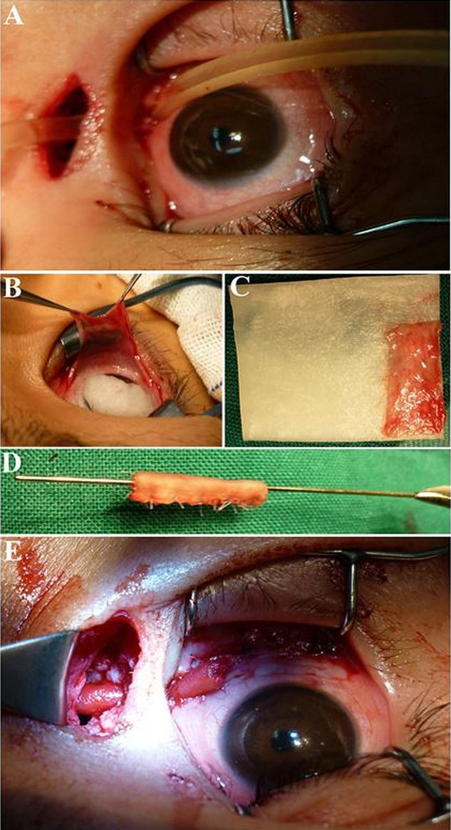
Figure 2.

The intraoperative pictures of the 9-year-old boy. There is a tunnel made from lacrimal caruncle through lacrimal sac area (A). The conjunctiva was harvested from inferior fornix (B). The conjunctiva was attached onto the oral biofilm (C). The oral biofilm was rolled into a tube shape with the conjunctiva being the internal lining (D). The newly-built duct was placed into the tunnel of lacrimal duct and fixed, with one end fixed onto the lacrimal caruncle and the other end fixed onto the nasal mucosa within the bony window (E).
