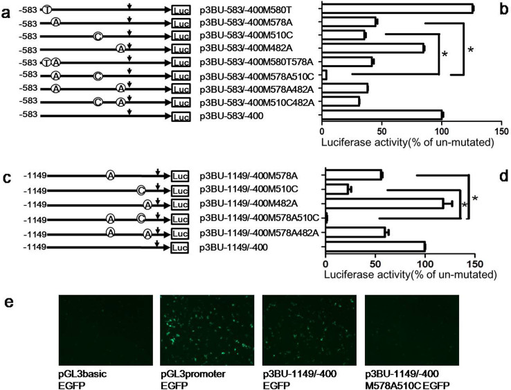
Figure 3. The cis-acting regions have a synergistic effect on activating BACE1 transcription.
(a)(c) Schematic diagram of plasmid constructs containing the BACE1 gene promoter with double nucleotide mutations with the plasmid p3BU-583/-400 or p3BU-1149/-400 as template. The horizontal line indicates the BACE1 promoter region; horizontal arrow indicates transcriptional direction and the downward arrow indicates the BACE1 transcriptional start site. Open circle with letter in it indicates mutation. The box LUC represents the coding sequence of the luciferase reporter gene. The numbers indicate the endpoints of each construct, with +1 as the adenine of the physiological translational start codon of the BACE1 gene. (b) (d) HEK293 cells were transiently transfected reporter plasmids. Firefly luciferase activity was measured 24 h after transfection, and Renilla luciferase activity was used to normalize for transfection efficiency. Data are shown as percentage of control samples transfected with wild-type plasmid. The values represent means standard error of the mean (n = 3–6). # or * P< 0.05 by analysis of variance (ANOVA) with the posthocNewmann-Keuls test when comparing with control. (e) HEK293 cells were transiently transfected EGFP reporter plasmids. Fluorescence was observed 24 h after transfection.

