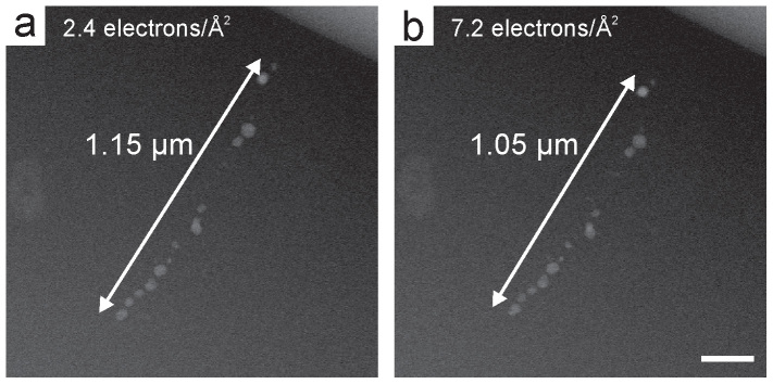
Figure 4. Electron beam damage of cells of M. magneticum by subsequent STEM acquisitions in the fluid cell.

(a) The first and (b) third HAADF-STEM images (cropped) of a cell. The cumulative electron doses and approximate magnetosome chain lengths are indicated in the images. The scale bar in (b) is 200 nm.
