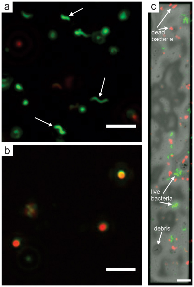
Figure 5. Composite fluorescence images of stained cells of M. magneticum prepared on a cover slip and in the fluid cell.
(a) Composite EGFP and rhodamine fluorescence images of cells of M. Magneticum concentrated approximately 20x from the original culture (same as fluid cell preparation). Several bacteria with intact membranes are denoted with white arrows. (b) Cells of M. magneticum killed with 10% isopropyl alcohol. (c) Composite EGFP, rhodamine, and brightfield optical image of bacteria in the fluid cell. The scale bar is 5 μm in (a) and (b) and 10 μm in (c).

