
Fig. 2.
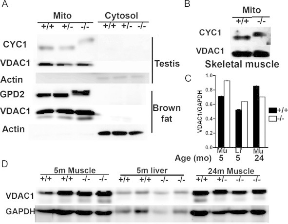
Expression of IMMP2L substrates in Immp2l mutant mice. (A) CYC1 and GPD2 expression determined by Western blot analyses in 3–5-month-old mutant mice. VDAC1 and β-actin were used as mitochondrial and cytosolic markers, respectively. (B) CYC1 expression in muscle mitochondria from 3 to 5-month-old mutant mice. (C) Densitometry comparison of VDAC1 expression in muscle and liver of control and mutant mice. Data were derived from panel “D”. Means±SEM are presented. Mu: skeletal muscle; Li: Liver. (D) Western blot analyses of VDAC1 expression between control and mutant mice using whole tissue lysates. GAPDH was used as a loading control.