
Fig. 1.
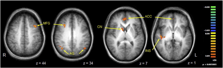
Contrast map of selected regions of BOLD activation that were significantly more active when vibrotactile stimuli were delivered to the symptomatic body part compared to the asymptomatic mirror region. Legend: anatomical images are derived from averages of T1-weighted scans from all participants and have been AC–PC aligned and transformed to Talairach space. MFG = middle frontal gyrus; AG = angular gyrus; CN = caudate nucleus; ACC = anterior cingulate cortex; INS = insula. See Table 1 for full list of activated regions. Per voxel cutoff of p < 0.005.