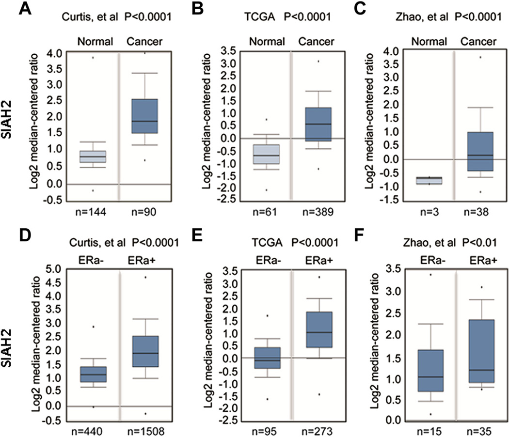
Fig. 1.
Expression of SIAH2 in breast cancer specimens and cell lines. (A) Data mining using the Oncomine program showing that SIAH2 is highly expressed in breast cancer specimens as compared with normal breast tissue. (B) SIAH2 expression also correlates with ER positivity in breast cancer. Each box represents multiple samples from one class. The whiskers indicate the normalized expression values including the maximum value, the 90th, 75th, median, 25th and 10th percentiles, and minimum values. The indicated numbers are cases of patient samples or normal tissue.

