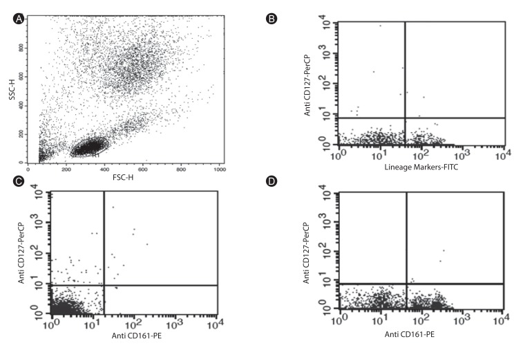
Fig. 3.
Flow cytometry analysis of the Lin-CD127+CD161+ innate lymphoid cells (ILCs) in the peripheral blood of the asthma patients compared with the healthy individuals. (A) Forward scatter/side scatter cannels diagram of peripheral blood cells shows gated area for lymphoid cells. (B) Flow cytometry analysis of the peripheral blood samples in which T cells, B cells, and natural killer cells are excluded as one population using the fluorescein isothiocyanate-conjugated anti-CD3, anti-CD19, and anti-CD56 antibodies. Lin-/CD127+ are seen in upper left area. (C) The diagram shows the Lin-CD127+CD161+ ILCs count in an asthma patient, which appears in upper right area. (D) Diagram of the Lin-CD127+CD161+ ILCs count in a normal control. These cells are in upper right area.

