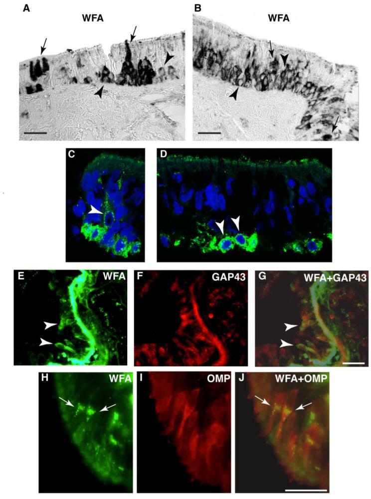
Fig. 1. Patterns of CSPG expression in the normal human OE.
In normal human OE, CSPG labeling using the broad spectrum marker WFA shows two distinct labeling patterns in OE cells: pericellular CSPG (p-CSPG) labeling (arrow heads), and cytoplasmic CSPG (c-CSPG) labeling (arrows). In A and B, light microscopy photomicrographs show examples of these two labeling patters. High resolution fluorescent confocal images of WFA labeled cells (green) and DAPI labeled nuclei (blue) confirm this pattern (C, D). In E-G, dual fluorescence labeling combining WFA with GAP43, prevalently expressed in immature ORNs, shows that p-CSPG+ ORNs correspond to GAP43+ORNs (arrow heads). In contrast, dual fluorescence labeling combining WFA with OMP, which typically labels mature ORNs, shows that c-CSPG+ ORNs correspond to OMP+ORNs (H-J; arrows). Together, these findings indicate that i) basal cells present with a pericellular pattern of CSPG/WFA; ii) immature ORNs maintain the p-CSPG expression pattern; iii) mature ORNs appear to transition to a prevalent cytoplasmic pattern of CSPG expression. Scale bar, 50 µm

