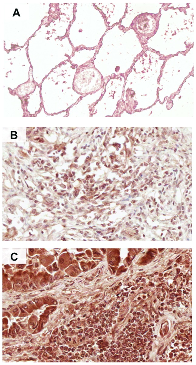
Figure 1.

Immunohistochemical detection of CXCR5 in lung cancer (LuCa) tissues. Representative figures of (A) non-neoplastic (n=8), (B) squamous cell carcinoma (SCC) (n=24) and (C) adenocarcinoma (AC) (n=54) lung tissues stained with anti-CXCR5 antibodies. Brown [3,3′-diaminobenzidine (DAB)] color shows CXCR5 staining. The images were captured using an Aperio ScanScope CS system with a 40× objective. Immuno-intensities of CXCR5 in each section were quantified using image analysis Aperio ImageScope v.6.25 software.
