Figure 6.
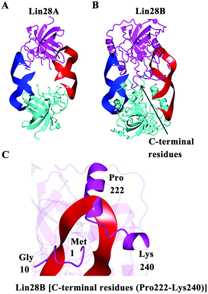
(A) Crystal structure of Lin28A:let7d complex (PDB:3TRZ). The complex consists of 2-fold symmetric Lin28A subunits (magenta/cyan) that coordinate the miRNA (red/blue) via interaction with the Lin28A CSD and ZNF domains. (B) Lin28B structure predicted from I-TASSER colored as in panel A. C-terminal residues of the putative Lin28B dimer are indicated by the arrow. (C) Zoomed-in view of the C-terminal residues (Pro 222-Lys 240) downstream of the ZNF2 domain as well as the N-terminal residues (Met1 to Gly 10) that would need to adopt a different conformation.

