
Figure 3.
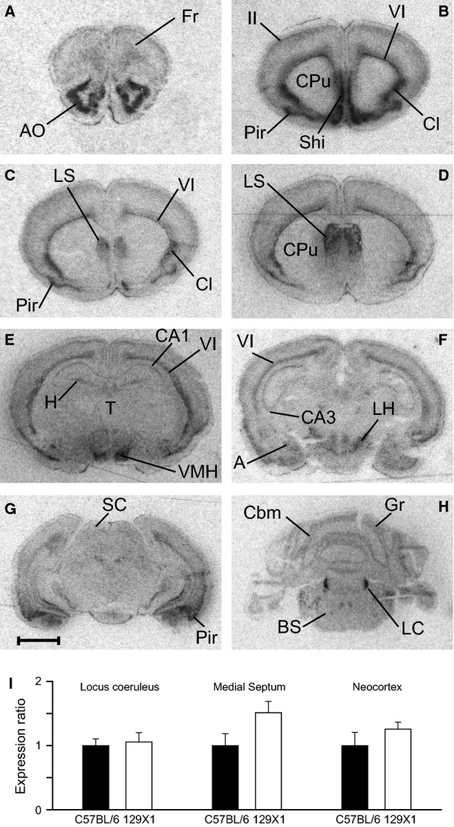
(A–H) In situ hybridization with a 33P-labelled oligonucleotide probe showing the distribution of Adra2a messenger RNA in coronal brain sections from 129X1 mice (n = 3). A, amygdala; AO, anterior olfactory nucleus; BS, brainstem; Cbm, cerebellum; Cl, claustrum; CPu, caudate-putamen; Fr, frontal cortex; Gr, granule cells; H, hippocampus; LC, locus coeruleus; LH, lateral hypothalamic area; LS, lateral septum; Pir, piriform cortex; SC, superior colliculi; Shi, septal hippocampal nucleus; T, thalamus; VMH, ventral medial hypothalamic area; ‘II’ and ‘VI’, layers of neocortex. Scale bar: 2 mm. (I) Expression levels of Adra2a mRNA assessed by real-time PCR do not differ (Student's t-test) between mouse strains (n = 6 for each strain) in the locus coeruleus (P = 0.77), the medial septum (P = 0.08) or the neocortex (P = 0.31).