Figure 1.

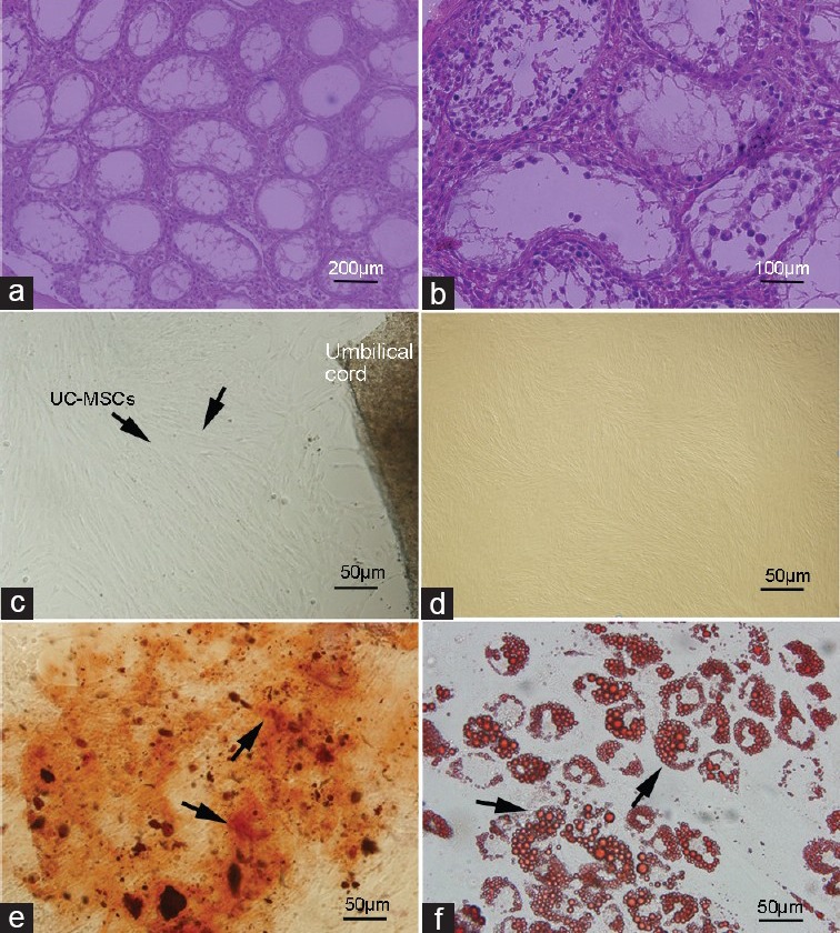
Histological examination of testis, cell culture and differentiation potential detection. Histological examination of testis sections from 5-week-old busulfan treated mice. Paraffin sections were stained with hematoxylin and eosin. 5 weeks after busulfan treatment, endogenous spermatogenesis was destroyed, and the testes of most are depleted in germ cells although they contain somatic cells and spermatogonia (a) ×200 (b) ×400. The umbilical cord samples were cut into small pieces, after 2 weeks, the cells migrate out of the tissue, and reach confluence. (c) Most of the cells were spindle-shaped and fibroblast-like. (d) At the third passage, adherent cells had the mesenchymal stem cell (MSC)-like phenotype. Differentiation capacity of human umbilical cord (HUC)-MSCs after expansion was detected. HUC-MSCs were induced to osteogenic (Von Kossa staining) (e) at days 21) lines and differentiate along adipogenic (Oil Red O staining) (f) at days 25). Multi-potent differentiation of HUC-MSCs was demonstrated.
