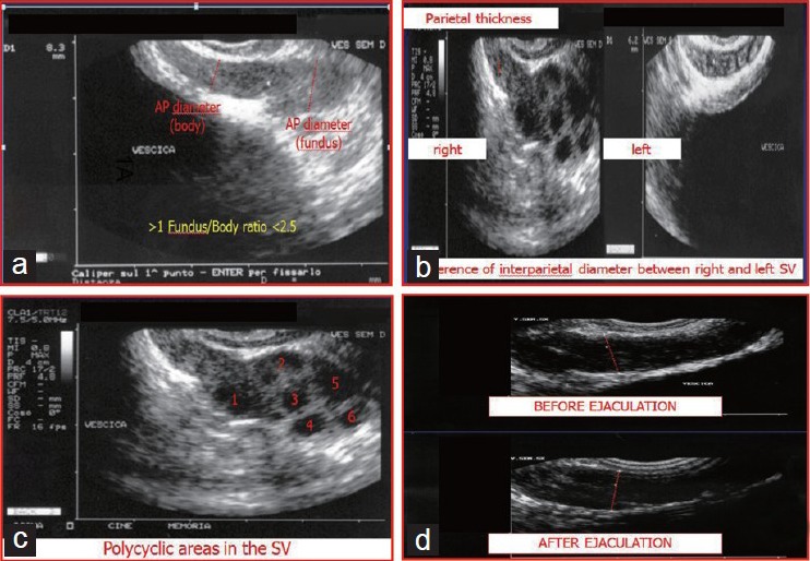
Figure 1.

Ultrasound parameters of the seminal vesicles (SVs) examined in the present study. (a) Body anteroposterior diameter (DAP), fundus DAP and fundus/body ratio; (b) Parietal thicknesses of the right and left SVs, difference in the parietal thicknesses between the right and the left SV and difference between the interparietal diameters of the right and the left SVs; (c) Number of polycyclic areas within both SVs; (d) Pre- and postejaculatory DAP differences.
