Fig. 5.
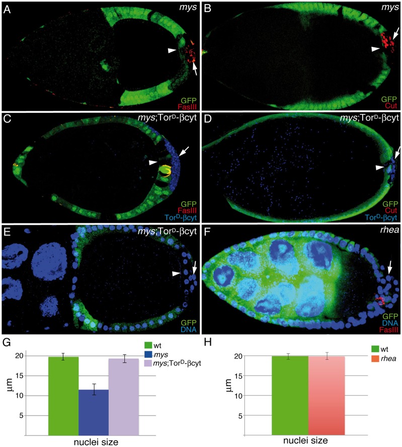
Integrin-mediated signaling is required for PFC maturation. Egg chambers were stained with TO-PRO-3 (E,F; blue), anti-GFP (A–F; green), anti-FasIII (A,C,F; red), anti-Cut (B,D; red) and anti-Myc (C,D; blue) to detect the TorD/βcyt chimeric integrin. (A,B) S9–S10 mosaic egg chambers showing abnormal expression of FasIII (A; red) and Cut (B; red) in mys PFCs located in ectopic layers (GFP−, arrow). (C–E) Ectopic expression of TorD/βcyt (blue) in these mys PFCs restores the normal expression of FasIII (C; red), Cut (D; red) and nuclear size (E,G). (F) FasIII expression is not affected in rhea PFCs located in ectopic layers (GFP−, arrow). (G,H) Quantification of the nuclear size of the indicated genotypes. The nuclear size of rhea PFCs is similar to that of wild-type (wt) follicle cells (H). In all cases, mys PFCs in contact with the germline (GFP−, arrowheads in A–E) behave as wild-type follicle cells. Data show the mean±s.d.

