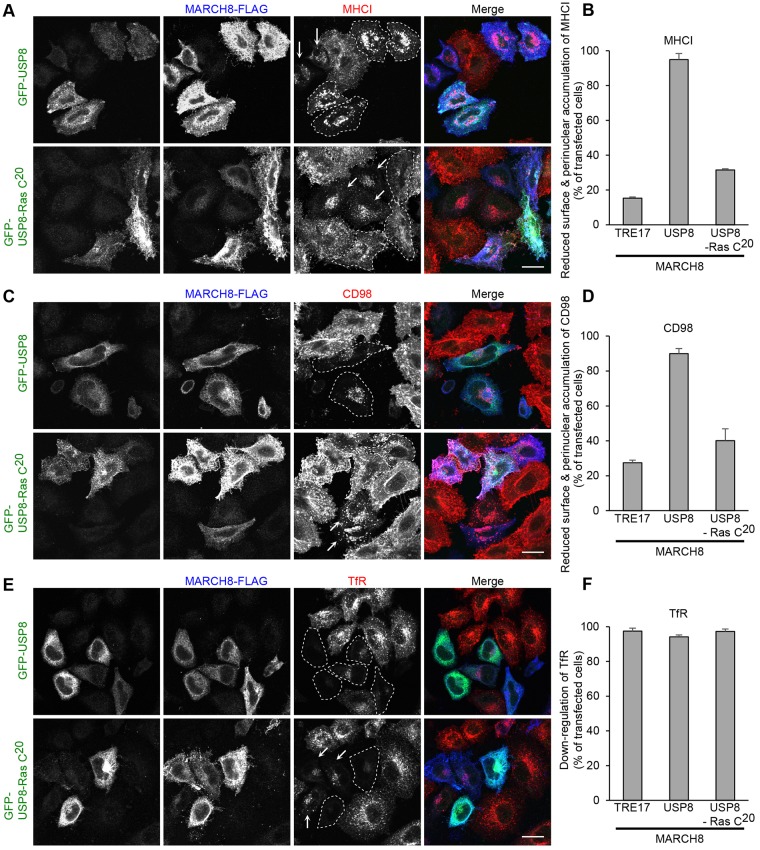
Fig. 6.
The USP8–Ras-C20 chimera counteracts the MARCH8-mediated targeting of CIE cargo proteins to late endosomes but not the downregulation of TfR. (A–D) HeLa cells were transfected with MARCH8–FLAG and GFP–TRE17 (not shown in A and C), and GFP–USP8 or GFP–USP8–Ras-C20. After 24 h, anti-MHCI (A,B) or anti-CD98 (C,D) antibodies were added to cells and treated as in Fig. 1. Cells were then fixed, and the internalized anti-MHCI (A,B) and anti-CD98 (C,D) antibodies were detected with anti-mouse-IgG antibody conjugated to Alexa Fluor 546 (red). GFP-tagged proteins were immunolabeled with chicken anti-GFP and anti-chicken-IgG antibody conjugated to Alexa Fluor 488 (green). MARCH8–FLAG was detected by rabbit anti-FLAG and anti-rabbit-IgG antibody conjugated to Alexa Fluor 633 (blue). The cell number was counted and plotted in B and D as in Fig. 1. Shown are mean±s.e.m. from three independent experiments. (E,F) HeLa cells were transfected with MARCH8–FLAG and GFP–TRE17 (not shown in E), GFP–USP8 or GFP–USP8–Ras-C20. After 24 h, cells were fixed and labeled with chicken anti-GFP, rabbit anti-FLAG and mouse anti-transferrin receptor, followed by immunofluorescence with anti-chicken-IgG antibody conjugated to Alexa Fluor 488 (green), anti-rabbit-IgG antibody conjugated to Alexa Fluor 633 (blue) and anti-mouse-IgG antibody conjugated to Alexa Fluor 546 (red). The cell number was counted and plotted in F as in Fig. 3. Shown are mean±s.e.m. from three independent experiments. In A, C and E, cells co-expressing MARCH8 and GFP-tagged proteins are outlined by dashed lines. Arrows indicate the cells expressing MARCH8 alone. Scale bars: 20 µm.

