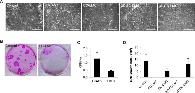
Figure 2.

Cell morphology, CFE, and cell growth rate of cultivated LECs from different culture methods. (A) Morphology of LECs from different culture methods. Dashed circles indicate areas containing epithelial-like cells. (B, C) The CFE of control and SS-LMC. (D) Cell growth rate of LECs from different culture methods. Scale bars: 100 μm.
