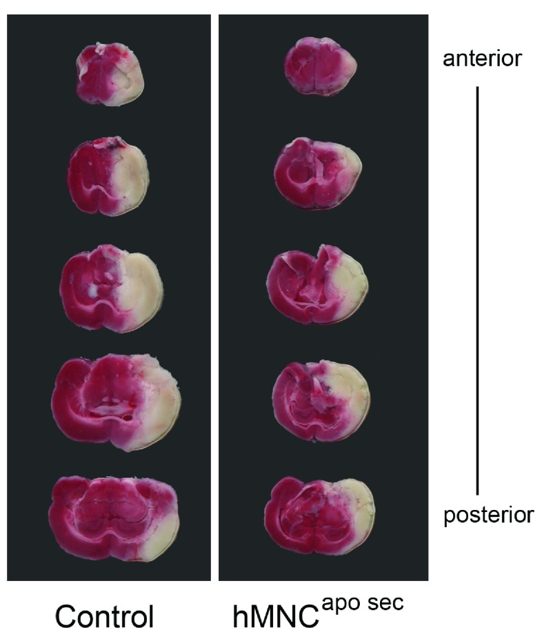
Figure 2. Representative brain slices of rats subjected to MCAO.
Brains were stained with a 2% solution of TTC forty-eight hours after MCAO. Animals received either treatment (in this representative scan: hMNC apo sec) or control medium, in this case, 40 minutes 24 hours after surgery. White areas indicate ischemic tissue while red areas stain for non-ischemic tissue. Animals treated with control medium (left image) had larger ischemic (=white) areas than animals treated with hMNC apo sec (right image).

