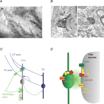
Figure 1. Cholinergic signalling modes in the thalamic reticular nucleus.

A, axonal arborizations with small diameter varicosities formed by cholinergic afferents in the rat TRN (Rt). Axons are immunostained for choline acetyltransferase (ChAT), the enzyme responsible for ACh synthesis. Scale bar, 20 μm. B, micrographs show two examples of axonal varicosities (labelled dark) formed by cholinergic axons in the TRN. Left image depicts an unmyelinated axon containing vesicles leading into a varicosity, without an obvious synaptic junction. Right image shows synaptic junction (outlined by arrows), formed onto a postsynaptic spine (sp). Scale bar, 1 μm. Images reproduced with permission from Parent & Descarries (2008). C, schematic diagram illustrates thalamic circuitry as well as possible targets of ACh signalling in the TRN. Cholinergic afferents (green) contact distal dendrites of TRN neurons and generate E–I cholinergic responses, probably mediated by ultrastructurally defined synapses (area outlined by square and shown in more detail in D). In addition, ACh released from en passant varicosities could act over larger distances and activate extrasynaptic receptors expressed on TRN dendrites, nAChRs expressed by thalamocortical (TC) axons or receptors expressed at presynaptic corticothalamic (CT) terminals. In turn, glutamate release from CT axons could modulate cholinergic signalling by acting on pre- or postsynaptic receptors at cholinergic synapses. D, schematic diagram illustrating the cellular mechanisms underlying cholinergic synaptic signalling in the TRN, based on the findings in Sun et al. (2013). ACh release activates postsynaptic ionotropic nAChRs (probably α4β2) and metabotropic Gi/o-coupled M2 mAChRs, which are expressed both pre- and postsynaptically. M2 mAChR activation in the postsynaptic membrane triggers the liberation of the βγ subunit complex, in turn leading to the opening of G protein-coupled inwardly rectifying K+ (GIRK) channels. Presynaptic M2 mAChRs are responsible for autoinhibition of ACh release, probably generated by βγ subunit complex-mediated inhibition of presynaptic Ca2+ channels.
