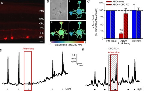
Figure 9. Adenosine inhibits light-evoked calcium responses from isolated ipRGCs in purified cultures.

A, immunolocalization of melanopsin in three ipRGCs located in the ganglion cell layer (GCL) of a retinal slice from an adult rat. The primary N-terminal melanopsin antibodies used here were employed in the immunopanning procedure to generate ipRGC cultures. Scale bar = 20 μm. B, light evoked a rise in internal calcium levels, measured as an increase in the fura-2 ratio, in isolated ipRGCs. Example pseudocoloured images of fura-2 fluorescence ratios in (i) a cultured ipRGC before (ii), immediately after (iii) and 50 s after (iv) exposure to 20 s light pulse. Scale bar = 25 μm. C, data summary for the experiments involving adenosine alone (n = 11) and adenosine plus DPCPX (n = 8). **P < 0.01, one-way repeated-measures ANOVA, Holm–Sidak. D and E, example traces showing that adenosine (D; ADO; 100 μm) reversibly inhibited the light-evoked calcium signals, and ADO's effect could be blocked by adding the A1 receptor antagonist DPCPX (E; 10 μm) to the bath. Stimulus was blue light (470 nm; 3.9 × 10¹⁵ photons s−1 cm−2).
