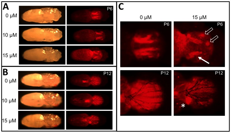
Figure 8. MeHg disruption of DLM muscle development.
(A, B) Mef2>RFP pupae reared on indicated concentration of MeHg to stage P6 (A) or P12 (B) and imaged by bright light and red fluorescence to reveal DLM morphology. (C) Close-up image of selected panel from A and B. The solid arrow indicates reduced DLM bundle size and defects in DLM bundle splitting with MeHg. Open arrows indicate displacement of attachment sites of DVM bundles. Asterisks (*) indicates failure of extension and anchoring of the DLM resulting in myofibers coalesced in a ball. The development of eyes and bristles appear unaffected by MeHg treatment.

