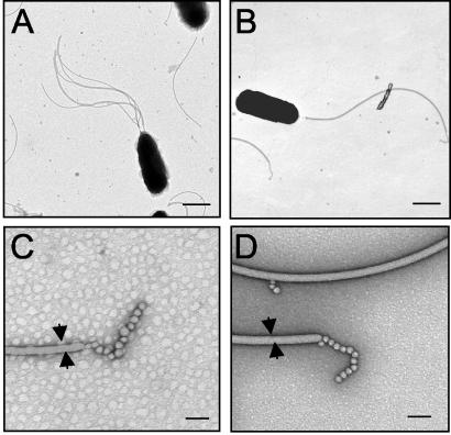
FIG. 5.
Transmission electron micrographs of V. fischeri cells in the mid-exponential growth phase. (A and B) Cells of wild-type strain ES114 (A) have more sheathed flagella than cells of the FlaA mutant DM143 (B). Bars = 500 nm. (C and D) At a higher magnification, the presence of a sheath, the diameter of the filament (arrows), and the presence of unknown structures at the distal end of the filament were similar for wild-type flagella (C) and FlaA mutant flagella (D). Bars = 50 nm.

