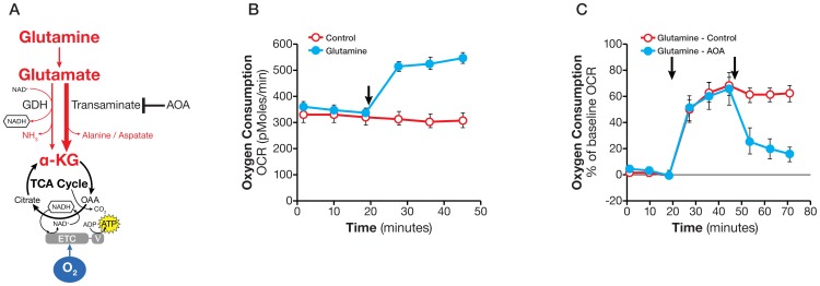
Figure 4. Assaying glutamine oxidation and demonstrating transaminase pathway activity.
A. Schematic illustration of biochemical pathway for glutamine oxidation in the mitochondria. B. Kinetic OCR response in SF188f cells to glutamine (4 mM). SF188f cells were plated at 20,000 cells/well in XF24 cell culture plates 24–28 hours prior to the assays. The assay medium was the substrate-free base medium. The OCR value was not normalized. A representative experiment out of three is shown here. Each data point represents mean ± SD, n = 4. C. OCR response (% of baseline) in SF188f cells to glutamine (4 mM) and AOA (100 µM). Glutamine-induced OCR reached 60% over the baseline (OCR at measurement 6 divided by that at measurement 3) while AOA addition reduced it to 20% (OCR at Measurement 9 divided by measurement 3). SF188f cells were plated at 20,000 cells/well in XF24 V7 cell culture plates 24–28 hours before the assays. The % OCR was plotted using measurement 3 as the baseline. The assay medium was the substrate-free base medium. A representative experiment out of three is shown here. Each data point represents mean ± SD, n = 4.

