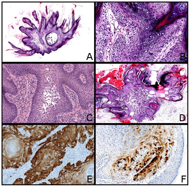
Figure 1. Histopathology and immunohistochemical findings of VP and SCC induced by vemurafenib.

(A) Typical VP with verrucous and papillomatous architecture covered by hyperkertosis (HE, x20). (B) Note the preeminent granulomatous layer with clear keratinocytes suggestive of an HPV infection (HE, x200). (C) VP with acantholysis (HE, x100). (D) VP with invasion of the superficial dermis (HE, x20). (E) Strong P16 positivity in a SCC. This tumor did not have any HPV (x100). (F) Heterogenous P16 expression in a VP (x100).
