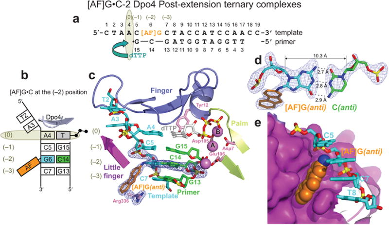
Figure 4.

Structure of the [AF]G•C-2 Dpo4 post-extension ternary complex. (a) Schematic of the expected pairing of the [AF]G-template with the 13-mer primer, ending with a 2′,3′-dideoxy-G, and added dTTP. (b) Schematic of the observed base pairing arrangement within the Dpo4 active site. (c) Structure of the active site. Simulated annealing Fo-Fc omit map contoured at 3σ level is colored in blue (2.0 Å resolution). (d) Watson-Crick base pair between the [AF]G(anti) and C14(anti) at the (–2) position. (e) Accommodation of the AF-moiety in a pocket on the surface of the little finger domain.
