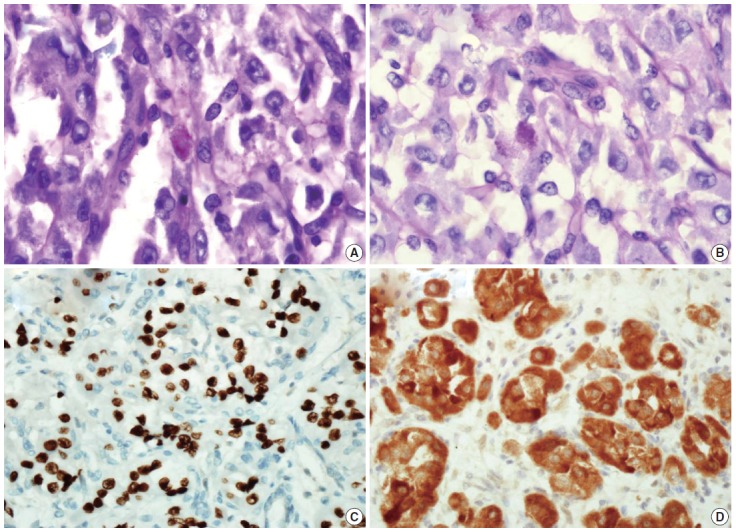
Fig. 3.

Periodic acid–Schiff (A) and periodic acid–Schiff with diastase (B) staining shows only poorly defined granules in the cytoplasm of the tumor cells. Immunohistochemically, the tumor cells are strongly nuclear positive for transcription factor E3 (C), positive for MyoD1 (D).
