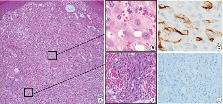
Fig. 2.
Histologic features of the first lesion. (A) The lesion is a poorly differentiated cellular tumor with focally infiltrating borders in the superficial dermis. (B) Most of the lesion shows solid proliferation of epithelioid endothelial cells. (C) Epithelioid endothelial cells are focally positive for CD34 immunohistochemical staining. (D) At the periphery, well-canalized vessels are observed. (E) Human herpesvirus-8 related antigen is negative.

