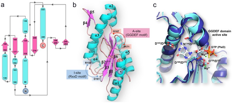
Figure 1. Domain architecture of tDGC.
(a) Topology of the GGDEF domain of tDGC. The N- and C- termini of the polypeptide chain are indicated. The β-strands are depicted by pink arrows and α-helices by blue tubes. (b) Location of the A- and I-site on the structure of tDGC: The loops bearing the “GGDEF” motif at the A-site and “RxxD” motif at the I-site are shown in red and blue respectively. (c) Superposition of the A site of tDGC (cyan) and PleD (dark blue)7. GTPαS-Mg2+, bound to PleD is shown as sticks and green spheres respectively. Residues involved in metal ion binding and base recognition (represented as sticks and labeled according to tDGC and PleD numbering schemes) are strictly conserved between the two proteins.

