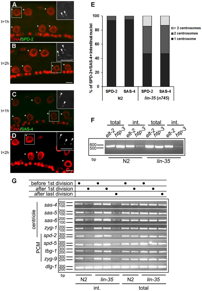
Figure 4. lin-35/Rb mutants undergo additional rounds of centriole duplication.
(A and B) lin-35/Rb mutant larvae were stained with DAPI (red) and anti-SPD-2 (green) to monitor centriole dynamics at the nuclear division. The panel (A) was obtained by staining animals approximately one hour after the nuclear division (t = 1 h), while (B) was acquired two hours after the division (t = 2 h). Asterisks indicate the intestinal nuclei and the arrowheads indicate SPD-2 foci. (C and D) lin-35/Rb mutant larvae were stained with DAPI (red) and anti-SAS-4 (green) to monitor centriole numbers after the nuclear division. The insets in (A–D) represent magnified views of regions highlighted by the white rectangles. Scale bar, 5 µm. (E) Quantification of SPD-2 or SAS-4 foci in intestinal nuclei in both wild type and lin-35/Rb mutants two hours after the first intestinal nuclear division. n = 75. (F) RT-PCR analysis of cell-specific transcripts from N2 and lin-35 (n745). elt-2 is intestinal specific, while htp-3 is expressed exclusively in the germ line. (G) The expression of spd-2, -5, zyg-1, -9, sas-4, -5, -6, tbg-1 and dlg-1 (control) [77] was quantified using RT-PCR from total or intestine-enriched mRNA from wild type (N2) or from lin-35 (n745) larvae before or after the first nuclear division, and 6–8 hours after the last nuclear division in lin-35 (n745) mutants. int., intestinal. bp, base pair.

