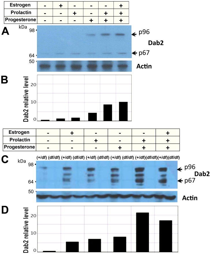
Figure 2. Hormonal induction of Dab2 expression in primary mammary epithelial cells.
Mammary epithelial cells were prepared from virgin wildtype control (fl/+) and Dab2 conditional knockout (df/df) mice inheriting Meox2-Cre. The cells were treated with 17 beta estradiol (1 nM), progesterone (1 µM), and prolactin (1 nM, or 50 ng/ml), individually or in combination for 4 days in culture. (A) Dab2 and beta-actin from primary mammary epithelial cells of virgin mice were analyzed by Western blot. (B) The signals of Dab2 proteins from the Western blot were quantified using NIH Image J software using beta-actin as normalization control. Relative values were plotted with the value of untreated cells defined as 1.0. (C) Dab2 and beta-actin from primary mammary epithelial cells isolated from pregnant mice were analyzed by Western blot. Dab2 protein was absent in cells from the conditional knockout mice. (D) The relative intensity of Dab2 proteins on Western blots was quantified using NIH Image J software, using beta-actin for normalization.

