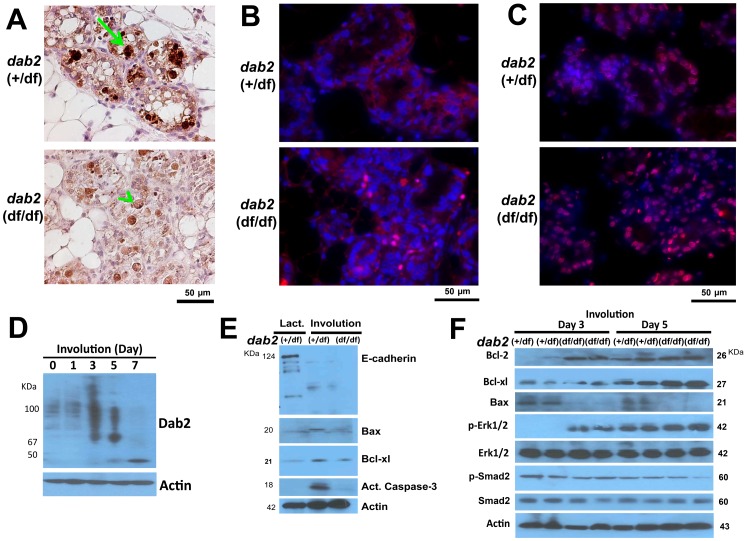
Figure 6. Delayed apoptosis of Dab2-deficient mammary epithelial cells during involution.
(A) The day 3 involuting mammary glands from control (dab2 heterozygous) and dab2 knockout (dab2 (f/df):Sox2-Cre) mice were analyzed. Apoptotic cell death in situ indicated by immunostaining for activated caspase-3, comparing wildtype (arrow) and Dab2 deficient cells (arrowhead). (B) A representative immunofluorescence microscopy staining for phospho-Erk1/2 in Dab2 heterozygous and knockout mammary glands on day 3 of involution. Phospho-Erk1/2 (red) overlaying on DAPI (blue) stainings are shown. (C) Immunofluorescence microscopy for Bcl-2 (red) and DAPI (blue) staining of day 3 involuting mammary glands. (D) Dab2 expression was determined by Western blot of tissue extracts of involuting mammary glands from dab2 heterozygous mice at 0, 1, 3, 5, and 7 days following forced involution. (E) Western blot analysis of E-cadherin, Bax, Bcl-xl, and activated caspase-3. Mammary protein extracts from heterozygous lactating (day 12) and forced involuting (day 3) mice were analyzed. (F) Western blot analysis of Bcl-2, Bcl-xl, Bax, phospho-Smad2, total Smad2, phospho-Erk1/2, and total Erk1/2 in protein lysates extracted from mammary glands following 3 and 5 days of forced involution. Two independent samples (duplicate) of each genotype from different mice are shown in the blot.

