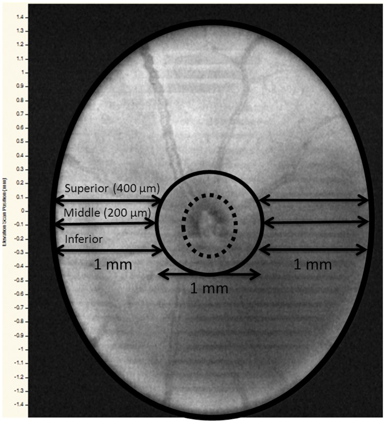
Figure 1. C57BL/6 mouse retina showing the reference points for SD-OCT and histological measurements.
Dotted circle – optic nerve head (ONH); black solid circle – 1 mm diameter central area surrounding the ONH; inferior, middle and superior reference points, each 200 µm apart, both to the left and the right of the ONH.

