
Figure 2. The fate of engrafted HGF-MSCs.
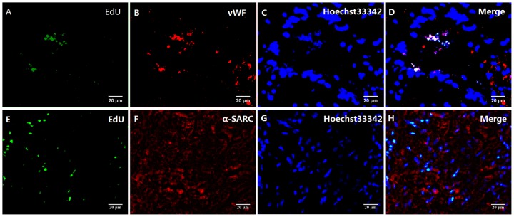
(A–D) Double-immunofluorescence staining for viable HGF-MSCs in the infaction border zone four weeks after transplantation. The merged fluorescence indicates colocalization of EdU and vWF, suggesting that the HGF-MSCs exhibited a tendency to differentiate into vascular endothelial cells. (E–H) There was a lack of evidence of cardiomyogenic differentiation based on the absence of α-SARC/EdU colocalization.