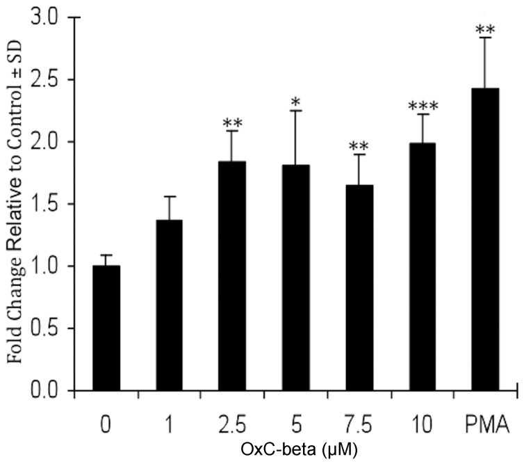
Figure 3. Phagocytosis in OxC-beta treated and LPS-stimulated THP-1 cells.
THP-1 monocytes were incubated with the indicated concentration of OxC-beta or DMSO control for 24 hours before being treated with LPS (15 ng/mL). Phagocytosis was evaluated 24 hours after LPS stimulation. Values represent fold changes relative to controls. PMA was used at 25 ng/mL. * p<0.05, ** p<0.02, *** p<0.002, Student’s t-test versus controls.

