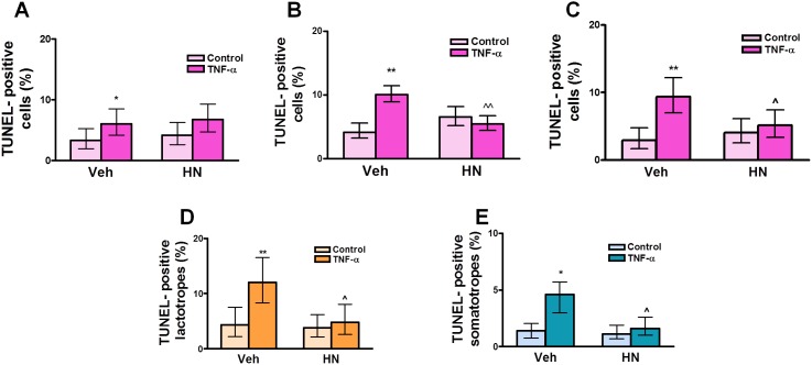
Figure 6. HN protects anterior pituitary cells from TNF-α-induced apoptosis in female rats.
Cultured anterior pituitary cells from OVX rats were incubated with 17β-estradiol (E2, 10−9 M) for 24 h and then with HN 2.5 µM (A), 5 µM (B), or 10 µM (C) for 2 h before adding TNF-α (50 ng/ml) for an additional 24 h. Apoptosis was assessed by the TUNEL method. In cells incubated with 5 µM HN, apoptotic lactotropes (D) and somatotropes (E) were identified by immunofluorescence. Each column represents the percentage ± CL of TUNEL-positive anterior pituitary cells (A, B, C, n≥1500 cells/group), lactotropes (D, n≥1600 cells/group) or somatotropes (E, n≥1800 cells/group). Data from at least two separate experiments were analyzed by χ2 test. *p<0.05, **p<0.01 vs respective control without TNF-α; ∧p<0.05, ∧∧p<0.01 vs respective control without HN.

