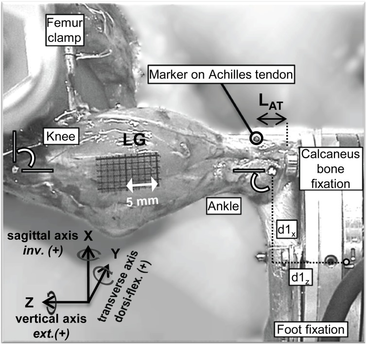
Figure 2. Lateral view of the rat left hindlimb in the set-up.
Ankle and knee joints were at 90° around the transverse axis. The femur was fixed and the foot was attached to the load cell using custom-made clamps. Anatomical landmarks for ankle and knee joint were aligned with the set-up’s rotational axes. A marker was placed on the posterior side of the Achilles tendon, which was used to assess length (LAT) and length changes of the distal part of the Achilles tendon. Positive ankle moments around the sagittal, transverse and vertical axis indicate inversion (inv.), dorsi-flexion (dorsi-flex.) and external rotation (ext.), respectively and are parallel to the x-, y- and z-axis of the load cell. In addition, d1x and d1z represent the distance from the center of the load cell to the center of the ankle joint in the x- and z-direction, respectively. LG: lateral gastrocnemius. Dotted vertical line represents the insertion site of the Achilles tendon.

