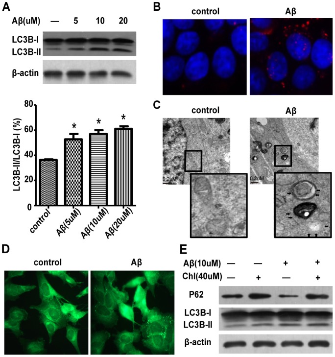
Figure 2. Aβ25–35 induces autophagy in SH-SY5Y cells.
(A) Western blot analysis of LC3 protein expression in SH-SY5Y cells treated with different doses of Aβ25–35 and quantification, β-actin was a loading control. (B) Immunofluorescence microscopy of punctate pattern of LC3 localization in SH-SY5Y cells treated with Aβ25–35. (C) Electron micrographs of SH-SY5Y cells treated with Aβ25–35 for 24 h. (D) Fluorescence microscopy of the formation of acidic vesicles after MDC staining in SH-SY5Y cells treated with Aβ25–35 for 4 h. (E)Western blot analyses of the protein expression of LC3 and p62 in cells treated with Aβ25–35 with and without chloroquine.

