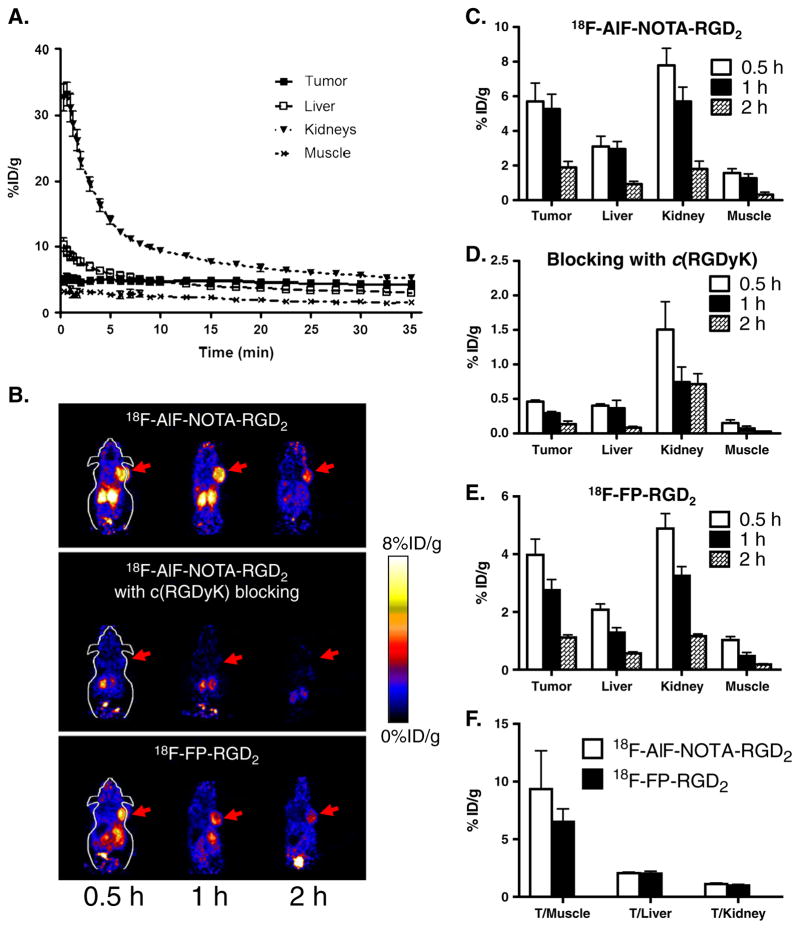
Fig. 5.
a Time-activity curves of tumor and major organs of athymic female nude mice bearing U87MG tumor from 35-min dynamic scans after intravenous injection of 18F-AlF-NOTA-RGD2 (~100 μCi/mouse, n=4). b Decay-corrected whole-body coronal microPET images of athymic female nude mice bearing U87MG tumor from a static scan at 0.5, 1, and 2 h after injection of 18F-AlF-NOTA-RGD2, 18F-AlF-NOTA-RGD2 with c(RGDyK) as blocking agent (10 mg/kg body weight), and 18F-FP-RGD2. Tumors are indicated by arrows. c–e MicroPET quantification of tumors and major organs at 0.5, 1, and 2 h after injection of 18F-AlF-NOTA-RGD2, 18F-AlF-NOTA-RGD2 with RGD as blocking (10 mg/kg body weight), and 18F-FP-RGD2, respectively. f Comparison of tumor to normal organ/tissue (muscle, kidney, liver) ratios of 18F-AlF-NOTA-RGD2 and 18F-FP-RGD2 at 2 h p.i.

