
Fig. 2.
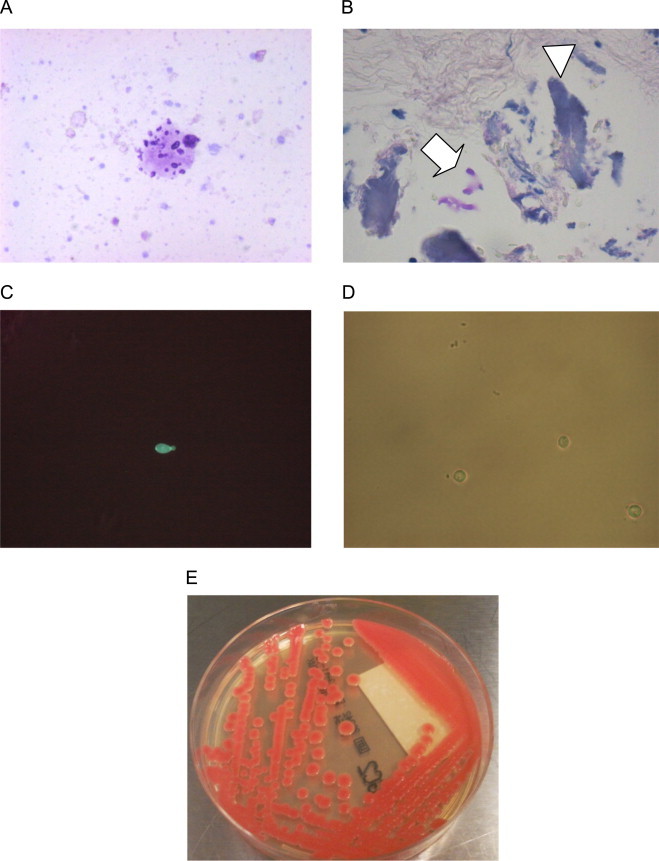
Cytology, histology and microbiology. (A) Fungal forms within proteinaceous material seen the cytological evaluation of the second CSF specimen obtained 7 days after the admission (Giemsa stain, 40×). (B) Focus of meningeal calcification (arrowhead) containing a few PAS (+) fungi (arrow), (PAS stain, 10×). (C) Fluorescence microscopy of biopsy specimen. (D) Optical microscopy of the yeast isolated after 4 weeks of incubation in liquid SAB (a capsule may be seen between the mother and the faint daughter cell). (E) The isolate of SAB agar.