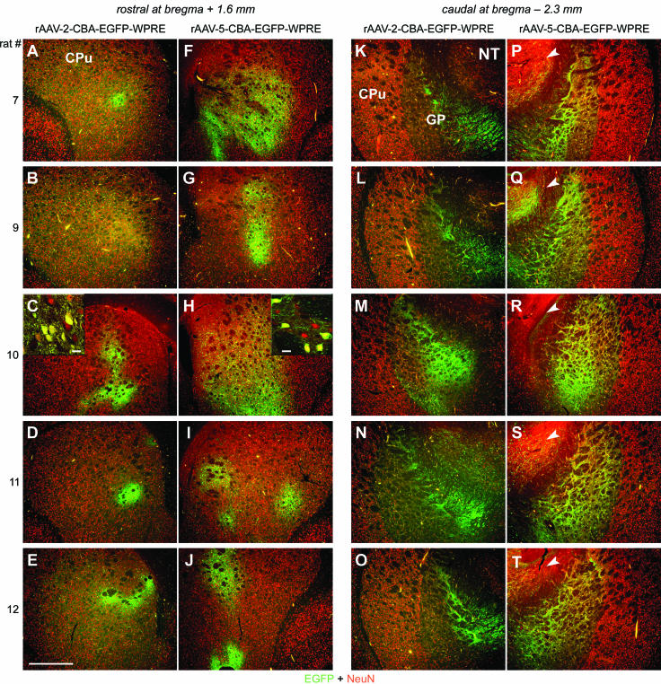
FIG. 1.

EGFP expression in the STR after striatal injection of rAAV-2 and rAAV-5. Coronal sections of the STR were analyzed for expression of EGFP (green) and NeuN (red) 28 days after virus injections. Representative two-color (merged) images from five animals are shown. (A to E) Within the rostral part of the STR, rAAV-2-mediated EGFP expression was locally restricted around the needle tract. CPu, caudate putamen. (F to J) In contrast, rAAV-5-mediated EGFP expression was widespread. (C and H insets) Confocal microscopy revealed that the majority of cells transduced within the STR by both serotypes were NeuN positive. Bars for insets, 25 μm. (K to T) Within the caudal part of the STR, EGFP expression was restricted to fibers (both serotypes) and a few positive cells (rAAV-2 [K to O]) in the GP. rAAV-5 transduced more cells within and outside of the GP (e.g., nucleus thalamus [NT; arrowheads in panels P to T]). Bar for panels, 500 μm.
