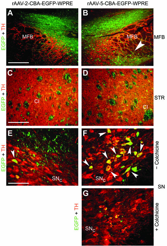
FIG. 5.

EGFP expression in the MFB, the STR, and the SN after injection of rAAV-2 and rAAV-5 into the MFB. Coronal sections of the MFB, the STR, and the SN were analyzed for EGFP (green) and TH (red) expression. Representative two-color (merged) images from two animals are shown. (A and B) EGFP-positive fibers and cells were present above and within the TH-positive area of the MFB after delivery of either serotype. However, only rAAV-5 transduced cells located on the ventral part of the MFB (B, arrowhead). Bar, 250 μm. (C and D) In the STR, EGFP-positive fibers were present in the CI of both hemispheres. Bar, 125 μm. (E and F) Occasionally, EGFP-expressing dopaminergic neurons were detected in the SNc after MFB injection of rAAV-2 (E, arrowhead), while more EGFP-transduced dopaminergic neurons were present after MFB delivery of rAAV-5 (F, arrowheads). In addition, a meshwork of EGFP-positive fibers surrounding the dopaminergic neurons was observed after MFB injection with either vector serotype. (G) Colchicine injection into the MFB prior to rAAV-5 delivery almost completely blocked EGFP expression in TH-positive cell bodies within the SNc. Bar for panels E to G, 62.5 μm.
