
FIG. 6.
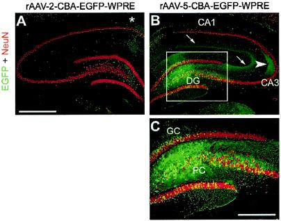
EGFP expression in the hippocampus after MFB injection of rAAV-2 and rAAV-5. Coronal sections of the hippocampus were analyzed for EGFP (green) and NeuN (red) expression. Representative two-color (merged) images from one animal are shown. A few EGFP-positive fibers and cells were present along the needle tract (which passed the hippocampus during MFB injection) after rAAV-2 injection (asterisk in panel A). In contrast, rAAV-5 transduced many granule cells (GC) and pyramidal cells (PC) as well as NeuN-negative cells within the DG (B and C; panel C is a magnification of the inset in panel B). In addition, EGFP-positive mossy fibers emanating from transduced GC were detected in their target area, the CA3 region (B, arrowhead), and EGFP-positive axons of transduced PC were present in the molecular layer of the hippocampus (B, arrows). Bar for panels A and B, 500 μm. Bar for panel C, 250 μm.