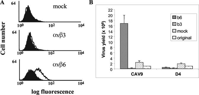
FIG. 3.

Flow cytometric analysis of CAV9 binding to SW480 cells expressing αvβ3 or αvβ6 and enhanced growth of CAV9 in cells expressing αvβ6. (A) Control cell samples were processed in the absence of virus (filled histogram). Virus binding (open histogram) was detected on SW480-αvβ6. Virus binding was not detected with SW480-mock or SW480-αvβ3 cells. (B) Levels (in PFU) of CAV9 and the RGD-less derivative D4 obtained when grown on monolayers of SW480-αvβ3 (b3), SW480-αvβ6 (b6), and SW480-mock (mock) cells for 72 h. The input virus inoculum is also shown (labeled “original”). The results are an average of duplicate measurements.
