Figure 1.

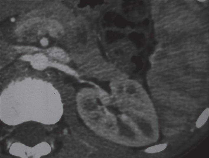
Computer tomography angiography demonstrates compression of the left renal vein between the aorta and superior mesenteric artery with dilation of the distal part of the left renal vein on the axial cuts

Computer tomography angiography demonstrates compression of the left renal vein between the aorta and superior mesenteric artery with dilation of the distal part of the left renal vein on the axial cuts