Figure 3.

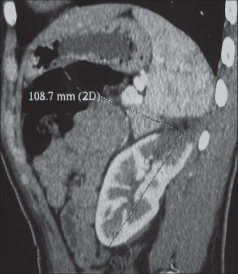
CT scan performed one month later shows the normal aspect of the kidney, except a little hematoma at the upper pole of the kidney and no compression of the left renal vein

CT scan performed one month later shows the normal aspect of the kidney, except a little hematoma at the upper pole of the kidney and no compression of the left renal vein